
Vision can change gradually, and small differences are easy to miss when both eyes work together. Simple checks at home can help you notice whether distance, near focus, color perception, central vision, or side vision seems different from usual. They are useful screening tools—not replacements for a comprehensive eye examination, prescription measurement, or diagnosis.
The five checks below require little or no equipment and include practical instructions, limitations, and signs that should prompt professional care. If a result concerns you, record what happened, repeat it under consistent conditions only when appropriate, and arrange an examination with an optometrist or ophthalmologist.
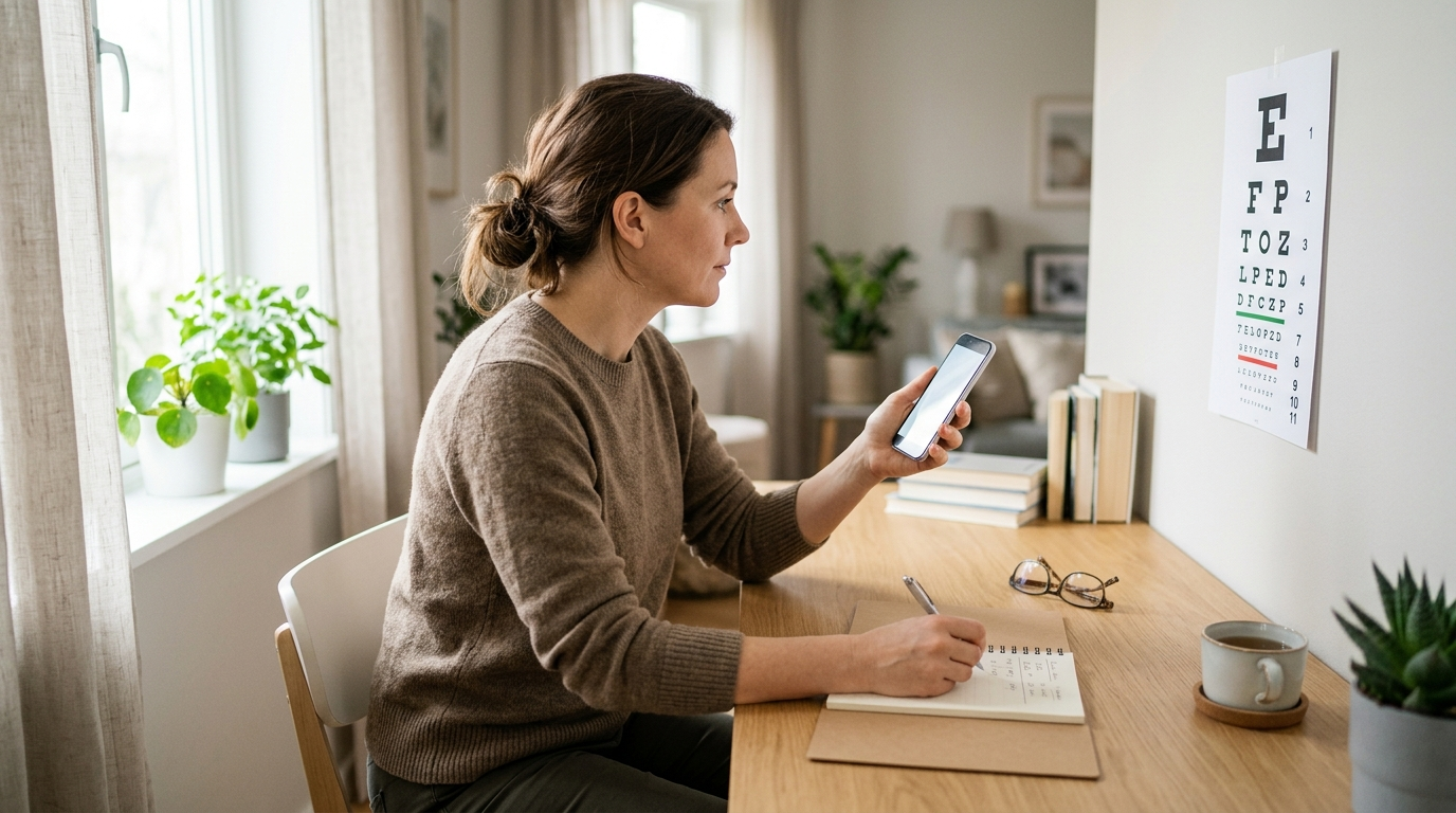
How to Prepare for a More Reliable Home Check
Consistency matters more than squeezing out a perfect score. Use good room lighting, remove glare from screens, and test one eye at a time while keeping your usual glasses on unless the instructions say otherwise. Do not press on a covered eye, and avoid testing when tired, dizzy, or experiencing sudden visual symptoms.
- Keep a tape measure or ruler available for distance checks.
- Use the same viewing distance each time; do not move closer to read a missed line.
- Have another adult help with side-vision testing for safety.
- Write down the date, eye tested, glasses or contact lens use, and result.
Home results can suggest that an appointment is worthwhile, but they cannot measure eye pressure, inspect the retina, assess the optic nerve, or identify every cause of blurred vision.
Do Not Rely on a Home Test for Sudden Symptoms
Some eye problems are time-sensitive and should not be monitored with an online chart or household test. Seek urgent medical advice for sudden vision loss, a curtain or shadow across vision, a new shower of floaters, flashes of light, severe eye pain, marked redness with nausea, or sudden double vision. These symptoms can require same-day evaluation.
- Emergency concern: abrupt loss of vision or a rapidly spreading field defect.
- Prompt appointment: persistent blur, headaches while reading, frequent squinting, or a noticeable difference between eyes.
- Routine monitoring: stable results with no symptoms and no known eye disease.
People with diabetes, glaucoma risk, macular disease, prior eye injury, or a history of eye surgery should follow their clinician’s examination schedule rather than substituting these checks.
Distance Visual Acuity Shows How Clearly You See Far Away
A distance acuity check is the familiar eye-chart test used to estimate how sharply each eye sees at a set distance. It can flag possible nearsightedness, farsightedness, astigmatism, an outdated prescription, or an imbalance between the eyes. Use a reputable printable chart or a calibrated digital chart, because an incorrectly sized chart can produce misleading results.
- Place the chart at the stated distance, commonly 10 feet or 20 feet depending on the chart design.
- Stand or sit upright with the chart at eye level and even lighting.
- Cover one eye gently without pressing on it, then read the smallest line where most characters are clear.
- Repeat with the other eye, first wearing your usual distance correction and then, if safe and useful, without it.
Results such as 20/20 are only meaningful when the distance and letter size are accurate. If one eye is substantially worse, you cannot read the expected line, or blur remains despite glasses, schedule a professional refraction and eye health assessment. Do not order new prescription lenses solely from a home result.
Near Vision Testing Reveals Reading and Screen-Focus Problems
Near vision can deteriorate even when distance vision seems normal. Difficulty with small print, shifting focus from a phone to a distant object, or needing to hold reading material farther away may reflect presbyopia, an uncorrected prescription, eye teaming difficulty, or another issue. A printed near-vision card is preferable to relying on a phone whose text can be resized.
- Use the card at its specified distance, often about 14 to 16 inches from the face.
- Test in comfortable reading light, without glare or very dim illumination.
- Check each eye separately, then both eyes together.
- Note whether words blur, double, move, or require unusually long focusing time.
A simple comparison can help you decide what to discuss:
| Pattern | Possible clue | Useful next step |
|---|---|---|
| Blur mainly up close | Presbyopia or near prescription need | Book a refraction, especially after age 40 |
| Intermittent blur with screens | Dryness, fatigue, or focusing strain | Use breaks and arrange an exam if persistent |
| Double or distorted print | Eye alignment or ocular-surface concern | Seek professional evaluation rather than stronger readers |
Use this check to start a conversation about reading glasses, occupational lenses, or screen habits—not to self-diagnose.
An Amsler Grid Check Can Flag Central-Vision Changes
An Amsler grid is a square pattern of lines with a central dot. It is commonly used for monitoring the central area of vision, particularly by people who have been advised to watch for macular changes. The test is most useful when performed regularly under similar conditions and compared with a prior result.
- Wear your usual reading correction and hold the grid at your normal reading distance.
- Cover one eye without pressing on it and stare directly at the center dot.
- Notice whether any lines look wavy, broken, missing, blurred, or unusually dark.
- Repeat with the other eye and record changes separately.
Do not let your eyes wander across the grid; doing so can hide a central defect. A new distortion, blank area, or sudden change should prompt a prompt call to an eye-care professional. An Amsler grid does not rule out retinal disease when it looks normal, and it cannot replace dilated retinal imaging or other ophthalmology assessments.
If you are researching the latest eye treatments or advanced ophthalmology options, bring dated grid notes and symptom timing to the consultation. They may help the clinician understand whether further imaging is appropriate.
Color-Perception Plates Help Identify Color-Vision Differences
Color-vision screening plates, such as number or shape plates viewed against colored dots, can reveal some inherited color-perception differences. They are useful for awareness, school or workplace discussions, and comparing whether one eye seems different from the other. They are not a complete test of color vision and may be affected by screen calibration, brightness, or color settings.
- Use a standardized test from a credible medical or educational source rather than a novelty quiz.
- View it in neutral daylight or consistent white light.
- Disable night mode, blue-light filters, and unusual display settings.
- Do not ask someone to name the expected answer before you respond.
Some color differences are lifelong and harmless, while a new or one-sided change deserves evaluation. A professional can use controlled lighting and additional tests to distinguish color-vision deficiency from problems affecting the lens, retina, optic nerve, or medication effects. If your work involves electrical wiring, transport, aviation, design, or safety signals, ask an eye-care provider which formal test your employer requires.
A Peripheral-Vision Check Screens for Missing Side Areas
Peripheral vision helps you detect movement and obstacles outside the point you are looking at. A basic confrontation check can identify an obvious difference between the two eyes, but it is not sensitive enough to exclude glaucoma or other field loss. Have a helper perform it, because testing yourself can lead to inaccurate head or eye movements.
- Sit facing the helper at roughly arm’s length, with your eyes level.
- Cover one eye while the helper covers the opposite eye, so each person uses the uncovered eye.
- Stare at the helper’s uncovered eye and keep your head still.
- Ask the helper to bring a finger or small target in from different sides and say when you first see it.
- Repeat in several directions and then test the other eye.
Missing a target repeatedly in one area, bumping into objects, or noticing a new dark section of vision warrants professional assessment. Formal visual-field testing uses controlled targets and maps the field more precisely. Never drive or operate equipment if your vision feels unreliable, even if this informal check appears normal.
What Home Screening Can and Cannot Tell You
These checks answer different questions, so no single pass or fail result represents overall eye health. A person may read a chart clearly while still having elevated eye pressure, retinal disease, dry-eye inflammation, or early glaucoma. Conversely, temporary blur from dryness or fatigue may make a healthy eye perform poorly on a particular evening.
| Check | Primary focus | Main limitation |
|---|---|---|
| Distance chart | Far-away sharpness | Accuracy depends on chart size and distance |
| Near card | Reading and close focus | Does not assess retinal or nerve health |
| Amsler grid | Central distortion | Not a full retinal examination |
| Color plates | Color discrimination | Screen and lighting can alter results |
| Side-vision check | Obvious field gaps | Less sensitive than clinical field mapping |
Use the results to choose a next step: monitor a stable, familiar pattern; arrange a routine exam for persistent concerns; or seek urgent care for sudden changes. Professional services may include refraction, slit-lamp examination, retinal evaluation, pressure measurement, and imaging.
Choosing Professional Care After an Unusual Result
Start with an optometrist or ophthalmologist who can perform a comprehensive examination and explain which finding matters. An optometrist is often an appropriate first contact for blurred vision, glasses, contact lenses, and routine screening; an ophthalmologist may be needed for suspected disease, surgical consultation, or complex treatment. A laser clinic near you may discuss LASIK or other laser eye procedures, but candidacy should follow a complete assessment—not an online test.
- Call the practice and describe the exact symptom, timing, and eye affected.
- Ask whether the visit includes a dilated exam, refraction, pressure measurement, and retinal imaging when indicated.
- Bring glasses, contact-lens details, medications, medical history, and your home-test notes.
- Request a written explanation of alternatives, risks, recovery, and total pricing before considering treatment.
Options such as monovision contact lenses or refractive surgery involve tradeoffs in depth perception, night vision, comfort, and adaptation. A consultation should explain those tradeoffs rather than promise a guaranteed result.
FAQs About Easy At-Home Eye Tests
Q: How often should I perform these tests?
A: There is no universal schedule. Occasional checks are reasonable for noticing change, while people with macular or other eye conditions should follow the monitoring plan provided by their clinician.
Q: Can an app replace an eye exam?
A: Apps may estimate visual acuity or provide reminders, but they generally cannot examine the retina, optic nerve, eye surface, or eye pressure. Treat app results as screening information only.
Q: Are online eye tests accurate enough to order glasses?
A: They may offer a rough estimate in controlled conditions, but accuracy varies with calibration and user technique. They should not replace an in-person examination, especially after a prescription change or when symptoms are present.
Q: What if one eye is consistently worse?
A: Arrange an eye examination, even if the difference is painless. A refractive imbalance may be straightforward to correct, but one-sided changes can also require evaluation of the cornea, lens, retina, or optic nerve.
Q: Can home tests detect glaucoma?
A: No. A home side-vision check can miss early field loss, and normal visual acuity does not exclude glaucoma. Risk-based professional screening is essential.
Turn a Home Result Into a Sensible Next Step
Use these five checks as a structured observation tool: test under consistent conditions, compare each eye separately, and record changes rather than chasing a perfect score. Persistent blur, distortion, double vision, or repeated difficulty should lead to a professional appointment. Sudden symptoms require urgent advice.
- Stable and symptom-free: keep routine eye examinations and protect comfort with regular screen breaks and good lighting.
- Repeatedly abnormal: book an optometry or ophthalmology visit and bring your notes.
- Sudden or severe change: contact urgent eye care immediately.
Once the cause is understood, you can compare appropriate options—including updated eyewear, contact-lens designs, medical treatment, or carefully evaluated LASIK alternatives—with much better information and realistic expectations.